Fundus examination and imaging - Dr. Mahmoud Hassaan.
A comprehensive evaluation of the retina and optic nerve using advanced diagnostic imaging to ensure early detection and management of retinal disorders.
 ● The fundus is examined at the retina clinic during a routine eye exam. Dilating eye drops are usually administered to ensure a clear view of the retina.
● Fundus examination with dilating eye drops is not routine but is performed in cases of symptoms, chronic diseases, or treatments that may affect the retina.
● Dilating eye drops can cause blurred vision for a period ranging from 6 to 24 hours or more, depending on the type of drops, their concentration, and the frequency of application. Therefore, it is advised not to rush into using dilating eye drops until vision testing and other eye function tests are completed, and to avoid driving until their effects have worn off.
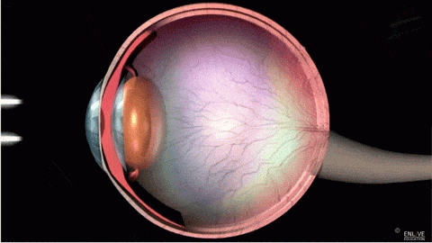
● The fundus examination includes examining the posterior surface of the lens, the vitreous humor, the retina, and the optic nerve.
● Fundus photography serves diagnostic purposes and is also important for documenting the visit, especially before and after surgical procedures.
● Fundus photography is performed using a specialized camera. Al Mashreq Eye Center is distinguished by its modern cameras with a wide field of view, providing a complete and clear view of the retina.
● Dr. Mahmoud Hassan advises patients to undergo fundus examinations and imaging if needed to avoid complications that could lead to vision loss.
● The fundus is examined at the retina clinic during a routine eye exam. Dilating eye drops are usually administered to ensure a clear view of the retina.
● Fundus examination with dilating eye drops is not routine but is performed in cases of symptoms, chronic diseases, or treatments that may affect the retina.
● Dilating eye drops can cause blurred vision for a period ranging from 6 to 24 hours or more, depending on the type of drops, their concentration, and the frequency of application. Therefore, it is advised not to rush into using dilating eye drops until vision testing and other eye function tests are completed, and to avoid driving until their effects have worn off.
● The fundus examination includes examining the posterior surface of the lens, the vitreous humor, the retina, and the optic nerve.
● Fundus photography serves diagnostic purposes and is also important for documenting the visit, especially before and after surgical procedures.
● Fundus photography is performed using a specialized camera. Al Mashreq Eye Center is distinguished by its modern cameras with a wide field of view, providing a complete and clear view of the retina.
● Dr. Mahmoud Hassan advises patients to undergo fundus examinations and imaging if needed to avoid complications that could lead to vision loss.
https://drmahmoud-hassaan.com/en/services/Eye-fundus-examination-imaging