Vitreous Removal and Retinal Detachment Repair
Vitreous removal and retinal detachment repair, including macular hole repair, using the finest surgical probes for precision.
 Our advanced surgical techniques for vitrectomy, also known as vitrectomy, along with the latest retinal detachment repair techniques, are designed to restore your vision. We specialize in complex cases, including macular hole repair, using the most precise and sophisticated surgical instruments available. This meticulous approach ensures the best possible results for preserving and improving your vision.
👁️ Vitrectomy and Retinal Detachment Repair
Retinal and vitreous surgeries are among the most delicate microsurgical procedures, requiring exceptional surgical skill and the latest global technology to guarantee vision restoration and eye wall stability.
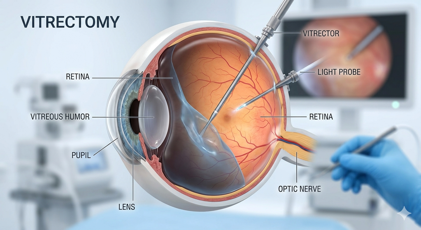
📌 What is a Vitrectomy?
It is a delicate surgical procedure in which the vitreous humor (the gel-like substance in the eye) is removed. This removal may be caused by complications of diabetes or penetrating injuries. The procedure allows direct access to the retina to repair any tears or holes, and then replace the vitreous humor with temporary or permanent prostheses to protect the eye.
---
🔍 Types of Retinal Detachment
Retinal detachment is medically divided into three main types, and the surgical treatment varies depending on the precise diagnosis:
1. Rhegmatogenous Retinal Detachment: This is the most common type and occurs as a result of tears or holes in the retina that allow vitreous fluid to leak underneath, separating it from the tissues that nourish it.
2. Tractional Retinal Detachment: This often occurs in patients with advanced diabetes, where scar tissue and membranes form on the surface of the retina, pulling it away from its normal position.
3. Exudative Retinal Detachment: This occurs as a result of fluid leaking under the retina without a tear and is often associated with infections or vascular diseases.
---
🏥 Clinic Preparation and Advanced Medical Examinations
The journey to successful treatment begins with accurate and rapid diagnosis; Therefore, the clinic is equipped with the latest global technologies for retinal health assessment:
- Optical Coherence Tomography (OCT): To image the retinal layers with micron-level precision and determine the size of leaks or holes in the macula.
- Fluorescein angiography (FA): To assess the efficiency of blood vessels and detect any retinal circulation deficiencies.
- B-Scan: To accurately assess the condition of the retina in cases of corneal opacities or acute vitreous hemorrhage.
-
🛡️ Operating Rooms and International Standard Equipment (ISO)
Surgeries are performed in a medical facility equipped according to the highest medical quality standards to ensure patient safety and world-class success rates:
- 7 fully equipped operating rooms: To accommodate all critical surgeries and eye emergencies at any time without waiting.
- ISO certification: Procedures are performed within a medical facility that strictly adheres to the ISO international standards for quality management and medical safety.
- Strict Sterilization Systems and HEPA Filters: Operating rooms are protected by a closed-loop system and equipped with high-efficiency particulate air (HEPA) filters, ensuring 99.97% air purification to prevent any intraocular infection during surgery.
---
🚀 Surgical Technology and Advanced Microscopes
We utilize a surgical system that represents the latest breakthrough in the history of eye surgery:
- NGENUITY® 3D Visualization System: We don't just look through traditional microscope lenses; we rely on the NGENUITY system, which allows the surgeon to perform the procedure while wearing 3D glasses and viewing a giant 4K screen. This system provides unprecedented depth of vision and ultra-high-resolution magnification of the finest details of the retinal membranes, while using very low light to protect the delicate retinal cells from photostress during the procedure.
- Advanced Surgical Microscopes: Supported by wide-field viewing systems for extremely precise examination of the retinal periphery, ensuring that all lateral tears are treated in the same session.
---
💧 Vitreous and Tamponade Alternatives
After retinal repair, an internal pressure device (tamponade) is needed to hold the retina in its natural position until it heals completely. These include:
- Temporary medical gases: These remain in the eye for several weeks, applying pressure to the retina. The body then absorbs them naturally and replaces them with the eye's natural fluids. A specific facial position is required after the procedure.
- Pure silicone oil: This is used as a long-term support in complex cases or severe detachments until the retina heals. It is removed later in a minor surgical procedure once the condition has stabilized.
Our advanced surgical techniques for vitrectomy, also known as vitrectomy, along with the latest retinal detachment repair techniques, are designed to restore your vision. We specialize in complex cases, including macular hole repair, using the most precise and sophisticated surgical instruments available. This meticulous approach ensures the best possible results for preserving and improving your vision.
👁️ Vitrectomy and Retinal Detachment Repair
Retinal and vitreous surgeries are among the most delicate microsurgical procedures, requiring exceptional surgical skill and the latest global technology to guarantee vision restoration and eye wall stability.
📌 What is a Vitrectomy?
It is a delicate surgical procedure in which the vitreous humor (the gel-like substance in the eye) is removed. This removal may be caused by complications of diabetes or penetrating injuries. The procedure allows direct access to the retina to repair any tears or holes, and then replace the vitreous humor with temporary or permanent prostheses to protect the eye.
---
🔍 Types of Retinal Detachment
Retinal detachment is medically divided into three main types, and the surgical treatment varies depending on the precise diagnosis:
1. Rhegmatogenous Retinal Detachment: This is the most common type and occurs as a result of tears or holes in the retina that allow vitreous fluid to leak underneath, separating it from the tissues that nourish it.
2. Tractional Retinal Detachment: This often occurs in patients with advanced diabetes, where scar tissue and membranes form on the surface of the retina, pulling it away from its normal position.
3. Exudative Retinal Detachment: This occurs as a result of fluid leaking under the retina without a tear and is often associated with infections or vascular diseases.
---
🏥 Clinic Preparation and Advanced Medical Examinations
The journey to successful treatment begins with accurate and rapid diagnosis; Therefore, the clinic is equipped with the latest global technologies for retinal health assessment:
- Optical Coherence Tomography (OCT): To image the retinal layers with micron-level precision and determine the size of leaks or holes in the macula.
- Fluorescein angiography (FA): To assess the efficiency of blood vessels and detect any retinal circulation deficiencies.
- B-Scan: To accurately assess the condition of the retina in cases of corneal opacities or acute vitreous hemorrhage.
-
🛡️ Operating Rooms and International Standard Equipment (ISO)
Surgeries are performed in a medical facility equipped according to the highest medical quality standards to ensure patient safety and world-class success rates:
- 7 fully equipped operating rooms: To accommodate all critical surgeries and eye emergencies at any time without waiting.
- ISO certification: Procedures are performed within a medical facility that strictly adheres to the ISO international standards for quality management and medical safety.
- Strict Sterilization Systems and HEPA Filters: Operating rooms are protected by a closed-loop system and equipped with high-efficiency particulate air (HEPA) filters, ensuring 99.97% air purification to prevent any intraocular infection during surgery.
---
🚀 Surgical Technology and Advanced Microscopes
We utilize a surgical system that represents the latest breakthrough in the history of eye surgery:
- NGENUITY® 3D Visualization System: We don't just look through traditional microscope lenses; we rely on the NGENUITY system, which allows the surgeon to perform the procedure while wearing 3D glasses and viewing a giant 4K screen. This system provides unprecedented depth of vision and ultra-high-resolution magnification of the finest details of the retinal membranes, while using very low light to protect the delicate retinal cells from photostress during the procedure.
- Advanced Surgical Microscopes: Supported by wide-field viewing systems for extremely precise examination of the retinal periphery, ensuring that all lateral tears are treated in the same session.
---
💧 Vitreous and Tamponade Alternatives
After retinal repair, an internal pressure device (tamponade) is needed to hold the retina in its natural position until it heals completely. These include:
- Temporary medical gases: These remain in the eye for several weeks, applying pressure to the retina. The body then absorbs them naturally and replaces them with the eye's natural fluids. A specific facial position is required after the procedure.
- Pure silicone oil: This is used as a long-term support in complex cases or severe detachments until the retina heals. It is removed later in a minor surgical procedure once the condition has stabilized.
https://drmahmoud-hassaan.com/en/services/vitrectomy-retinal-detachment